Δευτέρα 20 Οκτωβρίου 2014


xamhlh osfyalgia1

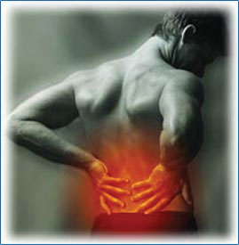
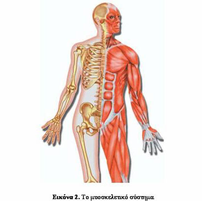
ΟΣΦΥΑΛΓΙΑ ΚΑΙ ΣΤΗΡΙΞΗ ΣΤΑ ΠΕΛΜΑΤΑFig1 heels

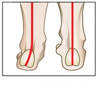
Όλοι οι ερευνητές συμφωνούν ότι η αρχιτεκτονική του άκρου πόδα σχετίζεται με την μορφολογία της λεκάνης, αλλά ένα ερώτημα που προκύπτει είναι: υπάρχει σύνδεση μεταξύ του τρόπου στήριξης στα πέλματα και την εμφάνιση οσφυαλγίας;

Ας υποθέσουμε ότι στην όρθια στάση ο αριστερός άκρος πόδας βρίσκεται σεπρηνισμό, που τυπικά προκαλείται από βλαισότητα στην υποστραγαλική άρθρωση (STJ), συνοδευόμενη από πτώση του σκαφοειδούς οστού. Ο δεξιός άκρο πόδας βρίσκεται σε υπτιασμό, όπου η υποστραγαλική άρθρωση είναι σε ραιβότητα, συνοδευόμενη από πτώση του κυβοειδούς οστού. Αυτός ο συνδυασμός πρηνισμού και υπτιασμού δεν επηρεάζει μόνο το φαινομενικό μήκος των ποδιών, αλλά ξεκινά μια μορφολογική διαταραχή που ταξιδεύει προς τα πάνω στην κινητική αλυσίδα, προκαλώντας στρέβλωση στο γόνατο, το ισχίο, τη λεκάνη και στις δομές της μέσης.
RotationPelvis
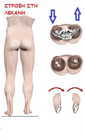
Καθώς ο πρηνισμός στον αριστερό άκρο πόδα προκαλεί έσω στροφή στον μηρό, ενώ ο υπτιασμός στον δεξιό άκρο πόδα προκαλεί έξω στροφή στον μηρό, το άτομο θα ωθείτο να βαδίζει πλαγίως σε κάθε βήμα του. Φυσικά το ιδιοδεκτικό σύστημα του οργανισμού δεν μπορεί να επιτρέψει κάτι τέτοιο, οπότε κάνει προσαρμογές. Μια από αυτές τις προσαρμογές σχετίζεται με τη θέση της κεφαλής του μηριαίου οστού στη κοτύλη.
Fig3 skeletalpelvis
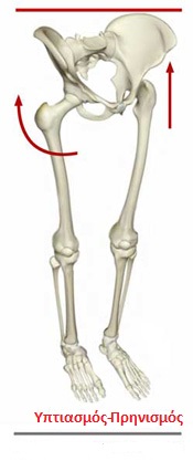
Στο αριστερό πόδι -που ο μηρός βρίσκεται σε έσω στροφή- ο αυχένας του μηριαίου οστού επίσης είναι σε έσω στροφή, γεγονός που ωθεί την κεφαλή προς τα πίσω ενάντια στην κοτύλη. Αντίθετα στο δεξιό πόδι -που ο μηρός βρίσκεται σε έξω στροφή- περιστρέφει τον αυχένα του μηριαίου, προκαλώντας ώθηση ενάντια στο πρόσθιο τμήμα της κοτύλης.
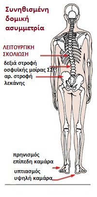
Με τη δεξιά μηριαία κεφαλή να ωθεί προς τα εμπρός και την αριστερή προς τα πίσω, τα οστά της λεκάνης ωθούνται σε αριστερή στροφή. Αυτό συμβαίνει γιατί -καθώς η ανυψωμένη κεφαλή του μηριαίου δεξιά οδηγεί την πρόσθια λαγόνια ακρολοφία προς επάνω και πίσω, περιστρέφοντας τη λεκάνη-, αντισταθμιστικά, η αριστερή πλευρά βυθίζεται. Ως εκ τούτου προκαλείται ασύμμετρη στροφή της λεκάνης, με αποτέλεσμα το μπλοκάρισμα στην οσφυϊκή μοίρα της σπονδυλικής στήλης. Δεν πρέπει να ξεχνάμε ότι η λεκάνη είναι η βάση στήριξης της σπονδυλικής στήλης. Η ασυμμετρία της θα προκαλέσει την προσαρμογή της σπονδυλικής στήλης σε μια σκολιωτική στάση, που θα οδηγήσει σε υπολειτουργικότητα και μυϊκή ανισορροπία.

Επικεντρώνοντας ο θεραπευτής την προσοχή του στη αποκατάσταση της συμμετρίας στην λεκάνη θα έχει, στην καλύτερη περίπτωση, ένα προσωρινό αποτέλεσμα, αλλά δεν θα έχει αποκαταστήσει το πρόβλημα του ασθενή.

CommonStructuralΟ πρηνισμός και ο υπτιασμός στον άκρο πόδα όχι μόνο μπορούν να προκαλέσουν στροφή της λεκάνης, αλλά μπορούν να οδηγήσουν σε μια αντισταθμιστική σκολιωτική στάση την οσφυϊκή μοίρα της σπονδυλικής στήλης, μεταφέροντας το πρόβλημα προς τα πάνω στη θωρακική και αυχενική περιοχή. Ο θεραπευτής επιβάλλεται να έχει μια σφαιρική προσέγγιση κατά τη διάρκεια της κλινικής αξιολόγησης, ώστε να αναγνωρίσει τον μηχανισμό πίσω από το πρόβλημα του ασθενή.
Επίσης, πολλές φορές διαπιστώνουμε πρηνισμό και στους δυο άκρους πόδες, με αποτέλεσμα αυξημένη οσφυϊκή λόρδωση, αυξημένη οσφύοϊερή γωνία και 'φρακάρισμα' στις αρθρώσεις (facets) μεταξύ του 4ο οσφυϊκού με 5ο οσφυϊκό σπόνδυλο και 5ου οσφυϊκού με 1ο ιερό σπόνδυλο. Στους ασθενείς αυτούς και οι δυο μηριαίες κεφαλές είναι τοποθετημένες προς τα πίσω, με αποτέλεσμα η λεκάνη να ωθείτε προς τα εμπρός και να αυξάνεται η λόρδωση στην οσφυϊκή μοίρα. Το αντίθετο συμβαίνει στη περίπτωση όπου διαπιστώνουμε υπτιασμό και στους δυο άκρους πόδες. Τότε προκαλείται ευθειασμός στην οσφυϊκή μοίρα της σπονδυλικής στήλης.


Όπως πολύ πειστικά ανέφερε ο Sir William Osler: «Για να μπορέσουμε να θεραπεύσουμε κάτι, πρέπει πρώτα να είμαστε σε θέση να το αναγνωρίσουμε»

static1


Βιβλιογραφία

1.
Zink G J. AN Osteopathic Structural Examination of the Soma. OsteopathicAnnals 7:12-19, 1979
2.
Previc F. A General Theory Concerning the Prenatal Origins of CerebralLateralizations in Humans. Psychological Review. Volume 98, 1991
3.
Geschwind N. Cerebral Lateralization. MIT Press, 1987


    ΠΡΟΒΛΗΜΑΤΑ ΠΟΥ ΠΡΟΚΑΛΟΥΝΤΑΙ ΣΤΟΥΣ ΧΡΗΣΤΕΣ COMPUTER
Μελέτες που έγιναν στις Η.Π.Α. το 1997 έδειξαν ότι 92 εκατομμύρια ενήλικες Αμερικάνοι πολίτες είναι χρήστες ηλεκτρονικών υπολογιστών. Περίπου το 40% έχουν υπολογιστή στο σπίτι τους, ενώ οι μισοί εργαζόμενοι χρησιμοποιούν υπολογιστή στον χώρο εργασία τους. Πολλές μελέτες έχουν γίνει κατά καιρούς για τις επιπτώσεις αυτής της χρήσης στο μυοσκελετικό σύστημα.
ΠΡΟΒΛΗΜΑΤΑ ΠΟΥ ΠΡΟΚΑΛΟΥΝΤΑΙ ΣΤΟΥΣ ΧΡΗΣΤΕΣ COMPUTER

Μελέτες που έγιναν στις Η.Π.Α. το 1997 έδειξαν ότι 92 εκατομμύρια ενήλικες Αμερικάνοι πολίτες είναι χρήστες ηλεκτρονικών υπολογιστών. Περίπου το 40% έχουν υπολογιστή στο σπίτι τους, ενώ οι μισοί εργαζόμενοι χρησιμοποιούν υπολογιστή στον χώρο εργασία τους. Πολλές μελέτες έχουν γίνει κατά καιρούς για τις επιπτώσεις αυτής της χρήσης στο μυοσκελετικό σύστημα.
computer users
Τελευταία έγινε μια μελέτη με σκοπό να καθορισθεί η συχνότητα περιπτώσεων εμφάνισης πόνου από την χρήση ηλεκτρονικών υπολογιστών στον χώρο εργασίας. Χρησιμοποιήθηκαν πάνω από 600 νεοδιορισμένοι υπάλληλοι, οι οποίοι χρησιμοποιούσαν υπολογιστή τουλάχιστον 15 ώρες την εβδομάδα. Τους ζητήθηκε να συμπληρώσουν ένα ημερολόγιο, όπου θα έπρεπε να αναφέρουν τις ώρες που χρησιμοποιούσαν τον υπολογιστή ημερησίως καθώς την παρουσία συμπτωμάτων πόνου στην περιοχή του αυχένα, στους ώμους αλλά και στα χέρια, για τρία χρόνια. Οι ερευνητές αναζήτησαν αυτούς που τα προσκληθέντα συμπτώματα απαιτούσαν φαρμακευτική αγωγή ή ήταν ψηλά στην κλίμακα αξιολόγησης του πόνου. Τα αποτελέσματα δημοσιεύτηκαν στο έγκυρο περιοδικό The American Journal of Industrial Medicine.
Περισσότεροι από το 50% των υπαλλήλων παρουσίασαν μυοσκελετικά προβλήματα τον πρώτο χρόνο της νέας εργασίας τους. Περίπου το 1/3 υπέφεραν από προβλήματα στην περιοχή του αυχένα και των ώμων, ενώ το ? από προβλήματα στα χέρια. Παράδοξα το Σύνδρομο του καρπιαίου σωλήνα, του περισσότερο γνωστού συνδρόμου της περιοχής ήταν σημαντικά χαμηλό, μόλις 1%. Μεγαλύτερη προδιάθεση παρουσίασαν οι γυναίκες ηλικίας 30 ετών και πάνω.
Αρχικά μπορεί αυτά τα συμπεράσματα να μας προκαλούν έκπληξη, αλλά να προσπαθήσουμε να δώσουμε μια εξήγηση την απάντηση θα μας την δώσει η διαπίστωση ότι η κακή στάση, η επαναλαμβανόμενη κουραστική εργασία σε συνδυασμό με το αδύνατο μυϊκό σύστημα προκαλούν αλλαγές στην διάταξη του σκελετού και κατά συνέπεια διαταραχή της δυναμικής ισορροπίας του σώματος.
Η θέση κάθε τμήματος του σώματος σε σχέση με τα υπόλοιπα ορίζει την στάση του σώματος. Σύμφωνα με τις δραστηριότητες του ατόμου η στάση αλλάζει συνεχώς. Τα οστά πηγαίνουν εκεί όπου οι μύες τα πηγαίνουν, ενώ συγκρατούνται εκεί όπου οι μύες τα συγκρατούν. Η ισορροπία σε αυτήν την δραστηριότητα προστατεύει το σώμα από τυχόν τραυματισμούς, είτε κατά την διάρκεια της εργασίας, είτε κατά την ξεκούραση.
Σωστή στάση σημαίνει οι αρθρώσεις να συγκρατούνται σε τέτοια θέση ώστε η επιβάρυνση που δέχονται να είναι ελάχιστη. Αντίθετα η λανθασμένη στάση προκαλεί μεγάλη επιβάρυνση αυξάνοντας την πιθανότητα τραυματισμού, όχι μόνο στην συγκεκριμένη περιοχή αλλά ακόμα σε άλλες λίγο πιο πάνω ή λίγο πιο κάτω από αυτήν. 
Το σώμα μας χαρακτηρίζεται από μία “αρμονία” κινήσεων.H μεγαλύτερη πρόκληση που είναι η υγεία, επιτυγχάνεται όταν όλα τα τμήματα του σώματος, είναι ελεύθερα να κινούνται σε αρμονική σχέση το ένα με το άλλο. Ο περιορισμός αυτής της συνεχούς εναλλαγής της κίνησης είναι υπεύθυνος για πολλές παθολογικές καταστάσεις. Για παράδειγμα η Σπονδυλική Στήλη πρέπει να είναι ισχυρή αλλά και λειτουργική, όχι μόνον γιατί προσφέρει στήριξη σε ολόκληρο το σώμα, αλλά γιατί προστατεύει τον Νωτιαίο Μυελό μέσο του οποίου επικοινωνεί και ελέγχει ο Εγκέφαλος ολόκληρο το σώμα. Οποιαδήποτε δυσλειτουργία στην σπονδυλική στήλη, μπορεί να επηρεάσει ολόκληρο το σώμα.
computer users1
Η στάση του σώματος αρχικά επηρεάζεται από την δύναμη της βαρύτητας. Στη συνέχεια η ένταση των καθημερινών δραστηριοτήτων προκαλεί σημαντική επιβάρυνση, που συχνά επιδεινώνεται από τις λανθασμένες θέσεις του σώματος, το αδύνατο μυϊκό σύστημα και τραυματισμούς. Το άμεσο αποτέλεσμα είναι φθορά στους μύες και τους συνδέσμους που συγκρατούν τον σκελετό. Τα βασικά συμπτώματα τότε είναι πόνος, περιορισμός της κίνησης και μυϊκή αδυναμία. Στην συνέχεια επηρεάζονται με την σειρά τους τα νεύρα της περιοχής με αποτέλεσμα φλεγμονές που προκαλούν ισχιαλγία, οσφυαλγία, αυχενικό σύνδρομο, πονοκεφάλους, πόνους στην πλάτη κ.α.. Ενώ η επιπλέον επιβάρυνση οδηγεί σε μη αναστρέψιμες βλάβες όπως είναι οι εκφυλιστικές αλλοιώσεις.
Πέρα από τις επιπτώσεις στην πάσχουσα περιοχή οι επιπτώσεις και στο υπόλοιπο σώμα είναι σημαντικές αν θεωρήσουμε το σώμα σαν ένα ενιαίο σύνολο. Για παράδειγμα η σκολίωση στην θωρακική μοίρα της σπονδυλικής στήλης επηρεάζει την αναπνευστική λειτουργία, λόγω περιορισμού της κινητικότητας του θώρακα, ακόμα επηρεάζεται το πεπτικό σύστημα και το κυκλοφορικό. Σε τελική ανάλυση διαταράσσεται η υγεία του ανθρώπου. 
Γενικά οι αιτίες που μπορούν να προκαλέσουν διαταραχή στην στάση, είτε μόνες τους, είτε σε συνδυασμό είναι:
• Επαναλαμβανόμενη κακή στάση στην επαγγελματική ενασχόληση.computer users2• Λανθασμένη στάση κατά την διάρκεια της ξεκούρασης.
• Ψυχολογικά ή συναισθηματικά προβλήματα, όπως η κατάθλιψη.
• Προβλήματα όρασης.
• Παχυσαρκία.
• Ακατάλληλα παπούτσια – διαταραχή της βάδισης.
• Αδύνατο μυϊκό σύστημα – διαταραχή της ισορροπίας στην βάδιση.
• Κακή διατροφή.
• Αναπνευστικά προβλήματα.
• Τραυματισμός μυών, τενόντων, συνδέσμών ή οστών.
• Σκολίωση.
Το ερώτημα είναι πώς μπορούμε να αντιμετωπίσουμε τις διαταραχές της στάσης, ώστε να αποκαταστήσουμε ή να βελτιώσουμε την υγεία του ανθρώπου.
Κάθε θεραπευτικό πρόγραμμα πρέπει να έχει σαν στόχο την λειτουργικότητα απαλλαγμένη από συμπτώματα. Η τεχνική "Manual Therapy", είναι η θεραπευτική προσέγγιση όπου ο θεραπευτής με την χρησιμοποίηση των χεριών του προσπαθεί να εντοπίσει, να αναγνωρίσει και τελικά χρησιμοποιώντας ήπιους χειρισμούς να αποκαταστήσει την ισορροπία στις πάσχουσες περιοχές του σώματος. Θεωρούμε το σώμα του ασθενή σαν ένα ενιαίο σύνολο με ψυχο-σωματική διάσταση, όπου η μορφή του εκφράζει την λειτουργικότητα του. Αυτή ακριβώς η μεθοδολογία της την κατατάσσει στις πλέον αποτελεσματικές και ασφαλείς θεραπευτικές μεθόδους με σχεδόν ανύπαρκτες επιπλοκές. Μέσα από την λεπτομερή αξιολόγηση του σώματος του ασθενή, οι πάσχουσες περιοχές μπορούν να αναγνωρισθούν και να αποκατασταθούν πριν προκαλέσουν μεγαλύτερο πρόβλημα. Στην συνέχεια εκπαιδεύουμε τον ασθενή να χρησιμοποιεί σωστά το σώμα του, ενώ τέλος γίνεται εκμάθηση ενός ατομικού προγράμματος ασκήσεων με σκοπό την ισχυροποίηση του μυϊκού συστήματος, ώστε ο ίδιος να αποκτήσει τον έλεγχο του προβλήματος (αυτοέλεγχος). 
Βασικό μέλημα του θεραπευτή είναι η βελτίωση της ποιότητας της ζωής του ασθενή, όπως αυτή εκφράζεται από την λειτουργικότητα του και την ψυχική του ισορροπία.
http://physio.gr/
Το " Δομικό " μοντέλο της μυοσκελετικής δυσλειτουργίας έχει άμεση και απόλυτη σχέση με την ανατομία , τις ανωμαλίες στην στάση και τις εκφυλιστικές αλλοιώσεις (π.χ. εκφυλισμός του μεσοσπονδυλίου δίσκου , οστεόφυτα , σκολίωση , κ.λ.π.) . 
Η παρουσία αυτών ενοχοποιείται σαν η βασική αιτία των συμπτωμάτων . pathiseis2 clip image002
Η παραπάνω θεωρία έχει στηριχθεί από την σημαντική ανάπτυξη των διαγνωστικών μέσων όπως η ακτινογραφία (X-RAY) ,η αξονική τομογραφία (CT SCAN) ,η μαγνητική τομογραφία (MRI) . 
Σκοπός της θεραπείας είναι η ανακατασκευή της δομής σύμφωνα με την τέλεια αρχιτεκτονική . Δυστυχώς όμως σε αρκετές περιπτώσεις οι κατασκευές αντιστέκονται στις δικές μας προσπάθειες .
Τον τελευταίο καιρό ενισχύεται η άποψη ότι ένα μεγάλο ποσοστό μυοσκελετικού πόνου και κατ' επέκταση της σωματικής δυσλειτουργίας που αυτό προκαλεί , έχει την ρίζα του στα μυοπεριτοναικά στοιχεία και όχι στους νευρικούς ή τους αρθρικούς ιστούς .
Ο Rosomoff και οι συνεργάτες του έχουν καταλήξει στο συμπέρασμα ότι κατά 90% και περισσότερο ο πόνος στη μέση οφείλεται στις αλλαγές στο μυοπεριτόναιο.
Ισχυρίζονται ότι η προέλευση του πόνου της μέσης δεν οφείλεται στην πίεση του νεύρου από εκφυλισμένο δίσκο ή από κήλη του δίσκου ( μία από τις περισσότερο δημοφιλείς θεωρίες ) , αλλά είναι το αποτέλεσμα του ονομαζόμενου σιωπηλού νεύρου και το πρόβλημα του πόνου στη μέση πρέπει να αντιμετωπιστεί σαν ένα μη χειρουργικό πρόβλημα , άσχετο με την νευρική πίεση . 
Το αποτέλεσμα της πίεσης στην νευρική ρίζα είναι η μείωση της αισθητικότητας και η απουσία των εκουσίων κινήσεων , όχι πόνος . 
Μόνον όταν γίνει άρση της πίεσης εμφανίζεται ο πόνος , παράλληλα με την βαθμιαία επιστροφή των εκουσίων κινήσεων .
Η εμπλοκή του μυοπεριτόναιου οφείλεται ,είτε σε βιοχημικούς παράγοντες, είτε σε απευθείας τραυματισμό του .
Ο Saal και οι συνεργάτες του ισχυρίζονται , ότι όταν ο δίσκος τραυματιστεί ή βρίσκεται σε διαδικασία εκφυλισμού , απελευθερώνει νερό και πρωτεογλυκάνες . 
Αυτό το υλικό στην συνέχεια , μετά από βιοχημική μεταμόρφωση μέσω της γλυκοξιλιώσης , αρχίζει μέσα από ένα μη ελεγχόμενο σύστημα , να συμπεριφέρεται σαν ξένο σώμα . 
Το αποτέλεσμα είναι μία φλεγμονώδης ανταπόκριση . Καθώς η συσσώρευση αυτής της « ξένης » πρωτεΐνης μέσα στον επισκληρίδιο χώρο συνεχίζεται , προκαλεί σημαντική αύξηση στα επίπεδα της φωσφωλιπάσης , που οδηγεί στην παραγωγή «επώδυνων» χημικών ουσιών , άρα βιοχημικά προκαλείται το αίσθημα του πόνου .
Ένας μικροτραυματισμός του μυός προκαλεί τη ρήξη κάποιων μυικών ινών και του ενδοπλασματικού δικτύου . Απελευθερώνονται ιόντα Ca ++ , με αποτέλεσμα τη συνεχή μυϊκή σύσπαση . Αυτό προκαλεί ισχαιμία και οίδημα από την συσσώρευση των ιόντων του μεταβολισμού. Έτσι δημιουργούνται ή γίνονται ενεργά τα trigger points . 
Αυτά προκαλούν πόνο που μπορεί να προβάλλει και σε άλλα μέρη του σώματος .Προκαλούν επίσης ελάττωση της μυικής ελαστικότητας και της μυικής ισχύος .
Ο μυς γίνεται ευάλωτος σε καινούργιο τραυματισμό , ο οποίος επαναλαμβάνει τον προηγούμενο κύκλο .Σε οποιοδήποτε σημείο της μυικής γαστέρας μπορεί να δημιουργηθεί ένα ενεργό TP , που μπορεί να συνοδεύεται και από άλλα δορυφορικά .
Ο τραυματισμός μπορεί να προκαλέσει και την δραστηριοποίηση του αντανακλαστικού τάσης ή μυοτατικού αντανακλαστικού σαν απάντηση στην διέγερση των μυικών ατράκτων από την απότομη διάταση των μυών .
Έχουμε δηλαδή ενεργοποίηση του γάμα νευρώνα.
Αυτή η τάση που αναπτύσσεται μεταφέρεται στην απονεύρωση είτε απευθείας , είτε από την κινητοποίηση του οστού που προσφύεται στην απονεύρωση .Έχει καθορισμένη διεύθυνση , εύρος και ένταση . Μεταφέρεται και στους υπόλοιπούς μυς που περιβάλλονται από την ίδια απονεύρωση , οπότε ενεργοποιείται εκ νέου και σε αυτούς το αντανακλαστικό τάσης , με την μεταφορά της τάσης και σε γειτονικές απονευρώσεις , κ.ο.κ. .
Το Κ.Ν.Σ. δέχεται ερεθίσματα που μπορεί να προέρχονται από οποιοδήποτε ιστό (περιτονία , μύ , αρθρικό θύλακο , μήνιγγα , σπλάχνα , δέρμα κ.λ.π. ) .
Ο νωτιαίος μυελός περιλαμβάνει αισθητικούς και κινητικούς νευρώνες όπου δέχεται αυτά τα ερεθίσματα . Σε περίπτωση όμως που τα ερεθίσματα ξεπεράσουν κάποιο όριο , τότε οι ηλεκτροχημικές εκκενώσεις αρχίζουν να επηρεάζουν παράλληλα μονοπάτια . Η διάχυση αυτών των ερεθισμάτων μπορεί να είναι μονόπλευρη ή ετερόπλευρη . Το αποτέλεσμα της υπερφόρτωσης του Κ.Ν.Σ. ορίζεται σαν Facilitated Segment ( FS ) - ΤΜΗΜΑ ΔΙΕΥΚΟΛΥΝΣΗΣ .
Σύμφωνα με τον UPLEDGER το FS μπορεί να εξηγήσει την ύπαρξη των παρακάτω προβλημάτων :
ΥΠΕΡΕΥΑΙΣΘΗΣΙΑ: Ελάχιστης έντασης ερεθίσματα μπορεί να προκαλέσουν υπερβολική ανταπόκριση .
ΥΠΕΡΧΕΙΛΙΣΗ : Τα ερεθίσματα μπορεί να γίνουν ασαφή και να διασκορπιστούν σε γειτονικά μονοπάτια προκαλώντας διιστάμενα συμπτώματα . Με αυτόν τον τρόπο προκαλείται ο προβαλλόμενος πόνος .
ΔΥΣΤΡΟΦΙΑ : Ένας χρόνιος ερεθισμός μπορεί να επηρεάσει τα συμπαθητικά και αυτόνομα μονοπάτια και να οδηγήσει σε τροφικές και μεταβολικές αλλαγές σε ιστούς που σχετίζονται με την μυοσκελετική δυσλειτουργία 
Το FS μπορεί από μόνο του να είναι πηγή προβλημάτων . 
Αν για παράδειγμα τραυματισμός του δικέφαλου προκαλέσει αυξημένα νευρικά ερεθίσματα , τα οποία μεταφέρονται με του τύπου Ια και ΙΙ νευρώνες , στο σπονδυλικό τμήμα Α5 και άλλοι μύες που συνδέονται με αυτό το τμήμα ( υπερακάνθιος , ελάσσων στρογγύλος , ανελκτήρ της ωμοπλάτης , ελάσσων θωρακικός , κ.λ.π. ) μπορεί να δεχτούν ερεθίσματα διάχυσης με αποτέλεσμα την ενεργοποίηση του γάμα νευρώνα και σε αυτούς . Αποτέλεσμα που οδηγεί σε αυξημένη σπαστικότητα και επιδεκτικότητα στην τάση . Άλλοι ιστοί ( δερματικοί υποδοχείς ,σπλάχνα , εγκεφαλικά κέντρα ) μπορεί επίσης να πέσουν σε αυτή την δοκιμασία άμεσα ή έμμεσα . 
Δεν πρέπει να ξεχνάμε το γεγονός ότι η περιτονία παίζει έναν έμμεσο ρόλο στον μεταβολισμό των οργάνων που περιβάλλει .Ακόμα ότι ο βασικός μεταβολισμός στα σπλάχνα εξαρτάται απευθείας από τις ρυθμικές κινήσεις της. Σε αυτό το τελευταίο μπορεί κανείς να προσθέσει τις ρυθμικές ή όχι κινήσεις του κινητικού συστήματος .
Η ελάττωση της υπερβολικής δραστηριότητας του γάμα νευρώνα καθώς και η απελευθέρωση των TPs είναι ο βασικός σκοπός για την ελάττωση της σπαστικότητας , ξανά τοποθετώντας τα αρχικά υποδείγματα της κίνησης , ενισχύοντας τον εκούσιο έλεγχο , βελτιώνοντας παράλληλα την μυϊκή ισχύ .
Η τεχνική S/CS φαίνεται να ανοίγει ένα παράθυρο για το Κ.Ν.Σ. να επανέλθει σε φυσιολογικές δραστηριότητες .
http://physio.gr/

Σάββατο 10 Μαΐου 2014


Γϊνεται ιδιοκτήτης

Οι μεγαλύτεροι ιστότοποι κοινωνικής δικτύωσης του κόσμου ρευστοποιούν εισόδημα της τάξεως αρκετών δισεκατομμυρίων δολλαρίων ετησίως ενεργοποιώντας δωρεάν χρήση των ιστοτόπων τους για τους χρήστες των.
Ο σκοπός του GlobAllShare δεν είναι απλώς να υλοποιήσει δωρεάν χρήση για τους χρήστες του, αλλά να τους κάνει να αισθανθούν την παγκόσμια κοινότητα ως δική τους και τοιουτοτρόπως να την διαμορφώσουν όπως καλύτερα νομίζουν. Γι' αυτό το GlobAllShare διασφαλίζει ιδιοκτησιακή κατάσταση σε κάθε χρήστη με την μορφή ελευθέρων μετοχών κατά την διάρκεια του σταδίου προοργανογράφησης.
Κάθε μέλος του GlobAllShare, που συμμετέχει στην κοινότητα κατά την διάρκεια του σταδίου προοργανογράφησης και συμβάλλει στην παγκόσμια ανάπυτξή του, μπορεί να λαμβάνει μετοχές του GlobAllShare συνεχώς και αδαπάνως και κάθε μήνα να λαμβάνει μερίσματα από το παγκόσμιο κέρδος κατ' αναλογία με τις μετοχές που έχει μετά που η παρούσα επιχείρηση ξεκινήσει.


 Είναι δωρεάν, οπότε δεν έχεις τίποτα να χάσεις. Είναι ένα ακόμα κοινωνικό δίκτυο όπως όλα όσα άλλα έχεις πιθανώς γραφτεί.

 για εγραφη πατιστε εδω
https://www.globallshare.com/el/2005860.html

Γραφτείτε και προσκαλέστε τους φίλους σας!!! Προσπαθείστε να πείσετε 5 φίλους σας να εγγραφούν μέσω του link που θα τους στείλετε και κερδίστε μια μετοχή εντελώς δωρεάν!!! Απλά προσκαλέστε τους φίλους σας και πείστε τους να προσκαλέσουν και αυτοί με τη σειρά τους τους φίλους τους!!

Για αυτούς που έχουν κάνει εγγραφή από τον παραπάνω σύνδεσμο και έχουν λογαριασμό στο Gas


 1 πιγενε στην αρχικη σελιδα

2 παστιστε το gas-vorld που ειναι στα αριστερα τισ σελιδας

3 διαβασε το κιμενο και μετα πατισε το κοκινο κουμπη τισ ενεργοπισεις

και μετα προσκαλεστε τουσ φιλους σας

Πάρτε την συμμετοχή σας από το παγκόσμιο κέρδος

Το GlobAllShare διαμοιράζεται το παγκόσμιο κέρδος του με τους χρήστες του, που έχουν συμβάλλει στην παγκόσμια ανάπτυξη και διάδοσή του κατά την διάρκεια του σταδίου της προοργανογραφήσεως.
Μόλις η επιχείρηση ξεκινήσει οι μέτοχοι του GlobAllShare θα λαμβάνουν κάθε μήνα τα μερίσματά τους από το 70% του παγκοσμίου κέρδους αναλόγως με τις μετοχές που κατέχουν, δηλαδή ο κάθε μέτοχος θα έχει πάγιο μηνιαίο κέρδος.
Όμως οι μέτοχοι του GlobAllShare δεν θα έχουν μόνο μηνιαίο μέρισμα αλλά θα μπορούν επίσης να πωλούν τις μετοχές των σε μια ονομαστική αξία, κερδίζοντας ένα άμεσο και μεγάλο ποσό με αυτόν τον τρόπο.

Αποκτήστε τις δικές σας μετοχές

Μπορείτε επίσης να αποκτήσετε τις δικές σας μετοχές στην κοινότητα του GlobAllShare απλώς κατά την διάρκεια του σταδίου της προοργανογράφησης. Το μόνο που χρειάζεστε είναι να προσκαλείτε φίλους και γνωστούς στον ιστότοπο του GlobAllShare.
Εάν έχετε προσκαλεσει 5 από τους φίλους σας με επιτυχία, θα πάρετε 1 μετοχή GlobAllShare αμέσως. Αλλά δεν μπορείτε απλώς να πάρετε μετοχές αφού καλέσει καποιος δικός σας γνωστούς, αλλά και μετά από κάθε περαιτέρω χρήστη που έχει προσκληθεί από τους φίλους σας, καθώς, άλλωστε μετά από γνωστούς φίλους σας πάρα πολύ, γιατί μέσα από 7 επόμενα επίπεδα μετά από κάθε ενας απο τους φίλους σας, ο οποίος συνειδητοποιεί 5 επιτυχημένες προσκλήσεις σας θα πάρετε επίσης 1 περισσότερο μερίδιο GlobAllShare.

συνεχές εισόδημα

Μετά τα συμφέροντά σας στην παγκόσμια κοινότητα του GlobAllShare μπορείτε να έχετε μια συνεχή μηνιαίο εισόδημα, από τη στιγμή που καταβάλλεται μέρισμα από την παγκόσμια κέρδη κάθε μήνα, κατ 'αναλογία του αριθμού των μετοχών που κατέχετε.
Εάν έχετε προσκαλεσει 5 από τους φίλους σας στην κοινότητα GlobAllShare με επιτυχία και ειπες τους φίλους σας να καλέσουν επίσης 5 από τους φίλους τους σε 7 επόμενα επίπεδα μετά από εσάς, τότε μπορεί να έχετε δικαίωμα στο μέρισμα εξής κάθε μήνα.
Επίπεδο 1 5 Προσκεκλημενοι 1 GAS Μετοχές
Επίπεδο 2 25 Προσκεκλημενοι 5 GAS Μετοχές
Επίπεδο 3 125 Προσκεκλημενοι 25 GAS Μετοχές
Επίπεδο 4 625 Προσκεκλημενοι 125 GAS Μετοχές
Επίπεδο 5 3 125 Προσκεκλημενοι 625 GAS Μετοχές
Επίπεδο 6 15 625 Προσκεκλημενοι 3 125 GAS Μετοχές
Επίπεδο 7 78 125 Προσκεκλημενοι 15 625 GAS Μετοχές
Σύνολο
19.531 GAS Μετοχές
Σύμφωνα με το παράδειγμα, μπορείτε να πάρετε συνολικά 19.531 GlobAllShare χαριστικών μετοχών, μετά την οποία θα πρέπει να καταβληθεί ένα μηνιαίο μέρισμα μετά την παγκόσμια έναρξη του εγχειρήματος. Αν, για παράδειγμα, θα καταβάλλονται μόνο $ 0.25 μερίσματος από την παγκόσμια κέρδη από ένα μήνα, θα πάρετε $ 5.000 το μήνα. Μπορείτε να πάρετε ακόμα και ένα μέρισμα μηνιαίο $ 0,5 ή 1,0 δολάρια μετά από κάθε μετοχές σας, έτσι μια μηνιαία κέρδη των $ 10.000 ή $ 20,000 μπορεί να παραχθεί εύκολα.
Μπορείτε να παρακολουθείτε τον αριθμό και την αξία των μετοχών, καθώς και το ποσό της μηνιαίας μερίσματος σας στο WebOffice μόνιμα.

Άμεσα χρήματα

Ως μέλος της κοινότητας GlobAllShare μπορείτε να πάρετε όχι μόνο ένα μηνιαίο εισόδημα, με τη μορφή μερισμάτων, αλλά και ένα μεγάλο χρηματικό ποσό, καθώς και από τη στιγμή που μπορεί να πωλήσετε τις μετοχές σας αμέσως
Μετά την παγκόσμια έναρξη του εγχειρήματος μπορείτε να πουλήσετε τις μετοχές σας στην ονομαστική τους αξία, κάνοντας έτσι ένα μεγάλο ποσό από τα χρήματα αμέσως. Ας πάρουμε ένα παράδειγμα: αν η ονομαστική αξία των μετοχών είναι $ 10 μετά την έναρξη της επιχείρησης και σύμφωνα με το παραπάνω παράδειγμα είστε ιδιοκτήτης 19.531 μετοχές GlobAllShare, μπορείτε να τα πουλήσει για συνολικά 195.531 αμερικανικά δολάρια, έτσι ώστε να κερδίσουν ένα μεγάλο ποσό αμέσως. Προφανώς, η ονομαστική αξία των μετοχών μπορεί εύκολα να φτάσει τα $ 20 ή $ 40 ανά μετοχή, ανάλογα με τον αριθμό των προσώπων που χρησιμοποιούν την κοινότητα portal GlobAllShare σε όλο τον κόσμο.
Θα πρέπει να σημειωθεί ότι οι χρήστες μπορούν να αποκτήσουν μετοχές GlobAllShare μόνο κατά τη διάρκεια της προ-οργάνωσης, σύμφωνα με την απόδοσή τους, ως εκ τούτου, σε παγκόσμιο έναρξη του εγχειρήματος μόνο αυτές θα έχουν τις μετοχές που συνέβαλαν στην ανάπτυξη του GlobAllShare.
Μετά την έναρξη της επιχείρησης θα εκδοθεί όχι οποιεσδήποτε άλλες μετοχές, έτσι μετοχές μπορούν να αγοραστούν από εκείνους αποκλειστικά οι οποίοι εμπλέκονται στην παγκόσμια δημιουργία του δικτύου και έλαβε μετοχών για τη συμβολή τους.

Κυριακή 6 Απριλίου 2014


What is a collarbone fracture?

A collarbone fracture is a break in the clavicle, the bone in your upper chest that connects your breastbone (sternum) to part of your shoulder blade (scapula).

What is the cause?

A broken collarbone can occur in several ways. You may fall on your outstretched arm and hand, you may fall on your shoulder, or you may be hit directly in the collarbone.

What are the symptoms?

You have pain and swelling at the area of the break. It is difficult to move your arm or shoulder. You may have heard a crack at the time of the injury.

How is it diagnosed?

A healthcare provider will need to examine the collarbone and do an X-ray to see if there is a break.

How is it treated?

To ease your discomfort, your collarbone may be immobilized in a "figure of 8" splint or brace that holds your shoulders back (as if you were standing at attention). Your arm may be put in a sling. Your provider will prescribe a pain medicine. Broken collarbones are very painful for the first few days.

Put an ice pack, gel pack, or package of frozen vegetables, wrapped in a cloth on the area every 3 to 4 hours, for up to 20 minutes at a time.

Only rarely will a broken collarbone need to be surgically repaired.

How long will the effects last?

Most broken collarbones are healed within 6 to 8 weeks. Your healthcare provider may take another X-ray to be sure that the bone has healed. You must be able to move your collarbone, shoulder, and arm without pain before you can return to your activities or sports. You can start rehabilitation exercises after your broken bone has healed and your provider says it is OK.

When can I return to my sport or activity?

Most broken collarbones are healed within 6 to 8 weeks. It is important for the collarbone to be fully healed before you return to your sport or activity so it doesn't break again. You must be able to move your collarbone, shoulder, and arm without pain. Your healthcare provider may take another X-ray to be sure that the bone has healed.

You can begin rehabilitation exercises after your broken collarbone has healed and after you've seen your provider.

How can I prevent a collarbone fracture?

Collarbone fractures are usually the result of accidents that cannot be prevented.

Comment, Like and Share with your Friends 

Multiple sclerosis (MS) is a disease in which the nerves of the central nervous system (CNS) degenerate. The CNS is made up of the brain and spinal cord. They process information from our environment and control voluntary muscle movements to allow the body to do certain things. This neural system works efficiently, unless there is a disease process affecting the pathways in the spinal cord and brain. Multiple sclerosis is one of the diseases that can affect these pathways and results in the destruction of myelin, a covering or insulation for nerves, that improves the conduction of impulses along the nerves and also is important for maintaining the health of the nerves. The demyelination (also known as plaques) disrupts the transmission of information in the CNS and leads to the symptoms seen in multiple sclerosis.

What Causes Multiple Sclerosis?
The cause of multiple sclerosis is still unknown. In the last 20 years, researchers have focused on disorders of the immune system and genetics for explanations, suspecting that a foreign agent such as a virus alters the immune system so that the immune system perceives myelin as an intruder and attacks it. Definitive proof of this viral theory is still lacking. However, the attack by the body's immune system on the neural tissues that it is supposed to protect is based on reasonable evidence. This attack is termed autoimmunity, thus making multiple sclerosis an autoimmune disease.

Much More Here: http://bit.ly/1cUxG9r


Cervical nerves are spinal nerves from the first seven vertebrae of the spinal cord. These seven vertebrae, called C1 through C7, begin at the base of the skull and make up the cervical spine in humans and most other mammals. There are eight cervical nerves, designated C1 through C8, with C1 through C7 emerging from above the cervical vertebra of the same number and nerve C8 emerging from below vertebra C7, the furthest from the skull. These nerves are connected to the muscles of the upper body and are among the most important in the nervous system.

Each cervical nerve innervates — stimulates — part of the upper body. Beyond the spine in the peripheral nervous system, these nerves branch, split, and intertwine. C1 through C4 form a structure in the neck called the cervical plexus, while C4 through C8, along with the first thoracic spinal nerve (T1), form the brachial plexus. Therefore, a particular cervical nerve can branch into multiple parts of the body, and a particular part of the body can be connected to more than one cervical nerve.

The head and neck are innervated by cervical nerves C1, C2, and C3. The thoracic diaphragm, a sheet of muscle across the lower rib cage essential to respiration, is innervated by C3, C4, and C5. Nerves C5, C6, C7, and C8, along with T1, split into a series of branches that innervate the arms, hands, and shoulders, as well as some muscles in the thorax.

Some of the nerves that branch off include the great auricular nerve, lesser occipital nerve, and transverse cervical nerve, which provide sensation on different parts of the head and neck, as well as the ansa cervicalis, which connects to the muscles of the neck. The phrenic nerve controls the diaphragm. Nerves connecting the cervical spine to the arms, shoulders, and hands include the radial nerve, ulnar nerve, and axillary nerve.

The cervical nerves are essential to sensation and motor control, and damage to them caused by disease or spinal injuries can result in partial or total loss of feeling and mobility in the upper body. It can also affect the autonomic nervous system in the torso, resulting in problems with digestion, respiration, and bowel and bladder control. Injuries at higher vertebrae generally cause greater impairment than injuries at lower vertebrae, and sufficient damage to the higher nerves can result in total quadriplegia — loss of use of all the limbs and the torso. The importance of the thoracic diaphragm to respiration means that a person can no longer breathe independently if the spine is severed above C3, although he or she can be kept alive by a respirator.

Article found here: http://bit.ly/ZMlSRY
Frozen shoulder, also called adhesive capsulitis, causes pain and stiffness in the shoulder. Over time, the shoulder becomes very hard to move.

Frozen shoulder occurs in about 2% of the general population. It most commonly affects people between the ages of 40 and 60, and occurs in women more often than men.

Anatomy
Your shoulder is a ball-and-socket joint made up of three bones: your upper arm bone (humerus), your shoulder blade (scapula), and your collarbone (clavicle).

The head of the upper arm bone fits into a shallow socket in your shoulder blade. Strong connective tissue, called the shoulder capsule, surrounds the joint.

To help your shoulder move more easily, synovial fluid lubricates the shoulder capsule and the joint.

Description
In frozen shoulder, the shoulder capsule thickens and becomes tight. Stiff bands of tissue — called adhesions — develop. In many cases, there is less synovial fluid in the joint.

The hallmark sign of this condition is being unable to move your shoulder - either on your own or with the help of someone else. It develops in three stages:

Freezing
In the"freezing" stage, you slowly have more and more pain. As the pain worsens, your shoulder loses range of motion. Freezing typically lasts from 6 weeks to 9 months.

Frozen
Painful symptoms may actually improve during this stage, but the stiffness remains. During the 4 to 6 months of the "frozen" stage, daily activities may be very difficult.

Thawing
Shoulder motion slowly improves during the "thawing" stage. Complete return to normal or close to normal strength and motion typically takes from 6 months to 2 years.
In frozen shoulder, the smooth tissues of the shoulder capsule become thick, stiff, and inflamed.

Cause
The causes of frozen shoulder are not fully understood. There is no clear connection to arm dominance or occupation. A few factors may put you more at risk for developing frozen shoulder.

Diabetes. Frozen shoulder occurs much more often in people with diabetes, affecting 10% to 20% of these individuals. The reason for this is not known.

Other diseases. Some additional medical problems associated with frozen shoulder include hypothyroidism, hyperthyroidism, Parkinson's disease, and cardiac disease.

Immobilization. Frozen shoulder can develop after a shoulder has been immobilized for a period of time due to surgery, a fracture, or other
injury. Having patients move their shoulders soon after injury or surgery is one measure prescribed to prevent frozen shoulder.

Symptoms
Pain from frozen shoulder is usually dull or aching. It is typically worse early in the course of the disease and when you move your arm. The pain is usually located over the outer shoulder area and sometimes the upper arm.

Doctor Examination
Physical Examination
After discussing your symptoms and medical history, your doctor will examine your shoulder. Your doctor will move your shoulder carefully in all directions to see if movement is limited and if pain occurs with the motion. The range of motion when someone else moves your shoulder is called "passive range of motion." Your doctor will compare this to the range of motion you display when you move your shoulder on your own ("active range of motion"). People with frozen shoulder have limited range of motion both actively and passively.

Imaging Tests
Other tests that may help your doctor rule out other causes of stiffness and pain include:

X-rays. Dense structures, such as bone, show up clearly on x-rays. X-rays may show other problems in your shoulder, such as arthritis.

Magnetic resonance imaging (MRI) and ultrasound. These studies can create better images of problems with soft tissues, such as a torn rotator cuff.
Treatment

Frozen shoulder generally gets better over time, although it may take up to 3 years.

The focus of treatment is to control pain and restore motion and strength through physical therapy.

Nonsurgical Treatment
More than 90% of patients improve with relatively simple treatments to control pain and restore motion.

Non-steroidal anti-inflammatory medicines. Drugs like aspirin and ibuprofen reduce pain and swelling.

Steroid injections. Cortisone is a powerful anti-inflammatory medicine that is injected directly into your shoulder joint.

Physical Therapy and Massage Therapy: Specific exercises will help restore motion. These may be under the supervision of a physical therapist or via a home program. Therapy includes stretching or range of motion exercises for the shoulder. Sometimes heat is used to help loosen the shoulder up before the stretching exercises. Below are examples of some of the exercises that might be recommended.

• External rotation — passive stretch. Stand in a doorway and bend your affected arm 90 degrees to reach the doorjamb. Keep your hand in place and rotate your body away from your hand. Hold for 30 seconds. Relax and repeat.

• Forward flexion — supine position. Lie on your back with your legs straight. Use your unaffected arm to lift your affected arm overhead until you feel a gentle stretch. Hold for 15 seconds and slowly lower to start position. Relax and repeat.

• Crossover arm stretch. Gently pull one arm across your chest just below your chin as far as possible without causing pain. Hold for 30 seconds. Relax and repeat.

Surgical Treatment
If your symptoms are not relieved by therapy and anti-inflammatory medicines, you and your doctor may discuss surgery. It is important to talk with your doctor about your potential for recovery continuing with simple treatments, and the risks involved with surgery.

The goal of surgery for frozen shoulder is to stretch and release the stiffened joint capsule. The most common methods include manipulation under anesthesia and shoulder arthroscopy.

Manipulation under anesthesia. During this procedure, you are put to sleep. Your doctor will force your shoulder to move which causes the capsule and scar tissue to stretch or tear. This releases the tightening and increases range of motion.

Shoulder arthroscopy.
In this procedure, your doctor will cut through tight portions of the joint capsule. This is done using pencil-sized instruments inserted through small incisions around your shoulder.
In many cases, manipulation and arthroscopy are used in combination to obtain maximum results. Most patients have very good outcomes with these procedures.

Recovery.
After surgery, physical therapy is necessary to maintain the motion that was achieved with surgery. Recovery times vary, from 6 weeks to three months. Although it is a slow process, your commitment to therapy is the most important factor in returning to all the activities you enjoy.
Long-term outcomes after surgery are generally good, with most patients having reduced or no pain and greatly improved range of motion. In some cases, however, even after several years, the motion does not return completely and a small amount of stiffness remains.

Although uncommon, frozen shoulder can recur, especially if a contributing factor like diabetes is still present.

Article First Seen Here: http://bit.ly/R6IoAZ

ΣΥΣΤΗΜΑ: ΙΔΙΟΔΕΚΤΙΚΟ

Οι αισθητηριακοί υποδοχείς για το ιδιοδεκτικό σύστημα βρίσκονται στους μυς και τις αρθρώσεις. Το σύστημα αυτό παίρνει πληροφορίες από τους μυς μας, τένοντες και συνδέσμους και μας δίνει μια αίσθηση του πού βρίσκεται το σώμα μας στον χώρο και τα όρια του σώματός μας. Μας επανατροφοδοτεί για την θέση του σώματος και την δύναμη και εύρος της κίνησης. Αυτό είναι που μας βοηθά να κάμψουμε το γόνατο στη σωστή γωνία για να σηκωθούμε από την καρέκλα, κατεβαίνοντας γλυκά από το πεζοδρόμιο και να χειριστούμε μολύβια, κουμπιά, κουτάλια και κόμπους (Anderson & Emmons 1996, Reisman & Hanchu 1992, SSI 1986). Το ιδιοδεκτικό σύστημα ερεθίζεται με την βαριά δουλειά ή την δουλειά με αντίσταση ή τις εκτάσεις στις αρθρώσεις. Όταν τραβούμε ή σπρώχνουμε κάτι βαρύ, οι αρθρώσεις απομακρύνονται λίγο ή συμπιέζονται, στέλνοντας πληροφόρηση στον εγκέφαλο για την δύναμη και το εύρος της κίνησης. Ο ερεθισμός του ιδιοδεκτικού συστήματος πιστεύεται ότι έχει ηρεμιστική επίδραση στους μαθητές εάν είναι υπερδιεγερμένοι και διεγερτική επίδραση στους υπο-διεγερμένους (Ayres 1979) – που σημαίνει, η ιδιοδεκτικότητα ομαλοποιεί την διέγερση από το υπέρ ή υπό ερεθιστικό επίπεδο σε ένα πιο θετικό επίπεδο.
Η σταθερή πίεση μπορεί να προστεθεί με το κρέμασμα, το έρπισμα κάτω από βαριές καρέκλες με σακουλάκια με φασόλια, ξάπλωμα κάτω από βαριές κουβέρτες και μεταφορά βαριών πραγμάτων. Οι εκτάσεις μπορούν να προστεθούν βάζοντας τους μαθητές να τραβούν κάτι βαρύ όπως ένα κομμάτι πλαστελίνης που έχει επικολληθεί στο θρανίο. Άλλος αποδεκτός τρόπος είναι να βάλουμε τον μαθητή να τακτοποιήσει τα βιβλία της βιβλιοθήκης χρησιμοποιώντας ένα βαγονέτο. Οι μαθητές μπορούν να στοιβιάσουν καρέκλες, ή να μαζέψουν τα τραπέζια ή να σφουγκαρίσουν το πάτωμα στην Τάξη. Η περιποίηση του κήπου είναι μια θαυμάσια και διασκεδαστική μέθοδος για να προωθηθεί η βαριά εργασία και να ενσωματωθεί στο πρόγραμμα μάθησης την ίδια στιγμή. Τα πηδήγματα σε μικρά τραμπολίνο προωθεί το ιδιοδεκτικό εισερχόμενο επιπρόσθετα με το αιθουσαίο εισερχόμενο. Η χρήση ανθεκτικών και ισχυρών χειρισμών (πχ lego, κύβοι ταιριάσματος, πλαστελίνη) αυξάνει τον ιδιοδεκτικό ερεθισμό στα χέρια. Το καθάρισμα του πίνακα, η μπουγάδα και το γέμισμα της μηχανής του χυμού μπορούν να προωθήσουν ιδιοδεκτικό ερεθισμό και προεπαγγελματική εκπαίδευση.
Οι άνθρωποι που έχουν προβλήματα σε αυτό το σύστημα έχουν την τάση να μην τοποθετούν καλά το σώμα τους σε σχέση με τα έπιπλα ή εργαλεία και να κάθονται στραβά στην καρέκλα ακόμη και εάν τα διορθώσουμε. Οι άνθρωποι με ιδιοδεκτικές δυσκολίες «έχουν δυσκολία να μετρήσουν τον βαθμό, την διεύθυνση και την αδιόρατη κρίση των κινήσεων που χρειάζονται για απαλές και συντονισμένες δράσεις. Μπορεί να χτυπούν πράγματα πάνω ή κάτω ή να μην πετυχαίνουν έναν στόχο» (κακή ποσόστωση δύναμης) (Reisman & Hanchu 1992 p.19, Anderson & Emmons 1996). Δεν μπορούν να πιάσουν μια μπάλα, να παίξουν ποδόσφαιρο (γιατί η μπάλα πηγαίνει πολύ γρήγορα για να την ακολουθήσουν τα μάτια) κλπ.
Συνήθως τα παιδιά με δυσκολίες σε αυτό το σύστημα είναι τα τελευταία που τα διαλέγουν για μια ομάδα σύμφωνα με τους Anderson & Emmons 1996. Χτυπούν το χέρι τους στα πράγματα όταν τα πλησιάζουν. Πολλές φορές όταν βάζουν ένα αντικείμενο σε ένα τραπέζι χτυπάνε το χέρι τους καθώς το τραβάνε, ή χτυπούν σε κόχες επίπλων. Μπορεί το πιρούνι να χάνει το στόμα και τρυπάει τα χείλη ή το μάγουλό. Μπορεί να προσπαθήσουν να πιάσουν το ποτήρι και να το πετάξουν κάτω. Αυτά τα κινητικά προβλήματα είναι πολύ ενοχλητικά και εξοργίζουν αφάνταστα το άτομο και το περιβάλλον του. Συνήθως τα γόνατά τους είναι χτυπημένα.
Κάποιες από τις αυτο–ερεθιστικές στρατηγικές που οι άνθρωποι με ιδιοδεκτική αναζήτηση αναπτύσσουν περιλαμβάνουν τα χτυπήματα των χεριών τους, τα πηδήματα, τα χοροπηδήματα, τα σκαρφαλώματα, τις κλίσεις προς ανθρώπους και αντικείμενα, τα συνεχή κουνήματα ενώ κάθονται και το βάδισμα στις μύτες των ποδιών (που αυξάνει την δύναμη των μυών των ποδιών) (Anderson & Emmons 1996, Reisman & Hanchu 1992). Αυτή η μη κοινωνικά αποδεκτή συμπεριφορά μπορεί να ελαττωθεί εάν η ιδιοδεκτική δράση προγραμματιστεί μέσα στην καθημερινή ρουτίνα. Ισχυρά εισερχόμενα στις αρθρώσεις είναι χαλαρωτικά όπως το σπρώξιμο, σκαρφάλωμα, άλματα και βαριά φυσική άσκηση. Σε πολλές περιπτώσεις, αυτό-τραυματιστικές συμπεριφορές μπορεί να αναπτυχθούν όπως το χτύπημα του κεφαλιού (που προωθεί στον εγκέφαλο πιο ισχυρά αιθουσαίες και ιδιοδεκτικές πληροφορίες). Το δάγκωμα είναι επίσης συχνό. Ερεθίζει μια ισχυρή ιδιοδεκτική αίσθηση στο σαγόνι (Reisman & Hanchu 1992).
Το ιδιοδεκτικό σύστημα εργάζεται με το αιθουσαίο σύστημα για να «χτίσει» τον μυϊκό τόνο. Ενώ το αιθουσαίο σύστημα σχετίζεται κυρίως με τον κύριο τόνο, το ιδιοδεκτικό σύστημα ρυθμίζει και οργανώνει γενικά υπόβαθρα του τόνου σε σχέση με όλο το κορμί. Οι άνθρωποι με αισθητηριακές δυσκολίες πρέπει να καταβάλλουν μεγαλύτερη προσπάθεια για να αρχίσουν και διατηρήσουν την κίνηση, έτσι κουράζονται πιο εύκολα και δείχνουν να είναι «τεμπέλικοι» και μη κινητοποιούμενοι όταν προσπαθούν να διατηρήσουν ενέργεια (Reisman & Hanchu 1992).
Η αιθουσαία αίσθηση δουλεύει και σε άλλες περιοχές κοντά στην ιδιοδεκτική αίσθηση. Το αιθουσαίο σύστημα βοηθά να σχεδιάσουμε μια κίνηση και το ιδιοδεκτικό να την εκτελέσουμε. Πρέπει να γνωρίζουμε πού βρίσκεται το σώμα μας στον χώρο για να μπορέσουμε να κινηθούμε γλυκά. Οι άνθρωποι με προβλήματα σε αυτά τα συστήματα συχνά τρακάρουν σε έπιπλα, πόρτες και άλλους ανθρώπους. Σχεδόν πάντα έχουν μελανιές κάπου στο σώμα τους ως αποτέλεσμα αυτού (Anderson & Emmons 1996).
ΔΥΣΛΕΙΤΟΥΡΓΙΕΣ ΙΔΙΟΔΕΚΤΙΚΟΥ ΣΥΣΤΗΜΑΤΟΣ
Πιθανές δυσλειτουργίες στην ιδιοδεκτική λειτουργία μπορεί να περιλαμβάνουν τα κάτωθι:
·Η κίνησή του είναι «σφιγμένη»
·Κουράζεται εύκολα, ιδίως όταν είναι όρθιο ή διατηρεί συγκεκριμένη στάση σώματος
·«Κλειδώνει» τις αρθρώσεις (πχ αγκώνες, γόνατα) για σταθερότητα (σφιγμένος)
·Δείχνει να έχει αδύναμους μυς
·Έχει αδύναμη σύλληψη με το χέρι
·Δεν μπορεί να σηκώσει βαρειά αντικείμενα (πχ είναι αδύναμο σε σύγκριση με τους συνομηλίκους)
·Θέλει να στηρίζει το σώμα του (ακόμη και σε δραστηριότητα)
·Περπατά στις μύτες των ποδιών
·Φτωχή αντοχή/κουράζεται εύκολα
·Δείχνει ληθαργικός (πχ δεν έχει ενέργεια, είναι νωθρός)
·Περνά την περισσότερη μέρα σε καθιστικό παιχνίδι (πχ κάνει ήρεμα πράγματα)
·Προτιμά το ήσυχο και καθιστικό παιχνίδι (πχ να βλέπει τηλεόραση, τα κομπιούτερς, βιβλία)
·Αποζητά περισσότερο παιχνίδια που τα παίζει καθιστός
·Προτιμά δραστηριότητες που τις κάνει καθιστός
·Διεγείρεται υπερβολικά κατά την κινητική δραστηριότητα
·Είναι συνέχεια «στην πρίζα»
·Αποφεύγει τις ήρεμες δραστηριότητες παιχνιδιού
·Το γράψιμό του είναι δυσανάγνωστο
·Έχει δυσκολία να μείνει μέσα στις γραμμές όταν βάφει ή γράφει
ΤΑΞΙΝΟΜΗΣΗ ΤΟΥ ΙΔΙΟΔΕΚΤΙΚΟΥ ΕΙΣΕΡΧΟΜΕΝΟΥ
ΔΙΕΓΕΡΤΙΚΟ:
Κεφάλι προς το έδαφος
Γρήγορη, ακανόνιστη, άρρυθμη
Ξαφνική απώλεια ισορροπίας
Αναπηδήσεις, πηδήγματα, κουνήματα
Γρήγορες κινήσεις κεφαλιού
ΗΡΕΜΙΣΤΙΚΟ:
Σιγανό, ρυθμικό λίκνισμα
Βαρειά εργασία

πηγη http://www.proseggisi.gr/

Προσοχή: ανοίγει σε νέο παράθυρο. PDF

Το παρακάτω πρόγραμμα έχει σχεδιαστεί για τη βελτίωση της ισορροπίας και της ιδιοδεκτικότητας (η ικανότητα να αισθανόμαστε ή να γνωρίζουμε που βρίσκονται οι αρθρώσεις μας στο χώρο, χωρίς να έχουμε οπτική επαφή).  Αυτό είναι σημαντικό γιατί βελτιώνουμε την ικανότητα μας να ρυθμίζουμε το κέντρο βάρους του σώματος μας σε κάθε αλλαγή θέσης ή σε κάθε κίνηση και να διατηρούμε τον έλεγχο.  Είναι επιστημονικά αποδειγμένο ότι βοηθούν στην πρόληψη τραυματισμών κι αποτελούν αναπόσπαστο κομμάτι της αποκατάστασης σε περίπτωση τραυματισμού του κάτω άκρου. Σε περίπτωση  τραυματισμού στην περιοχή πριν εκτελέσετε οποιαδήποτε από τις παρακάτω ασκήσεις πρέπει συμβουλευτείτε τον φυσικοθεραπευτή σας.

Συνήθως οι ασκήσεις ισορροπίας εκτελούνται 10 με 15 λεπτά την ημέρα χωρίς να αναπαράγουν συμπτώματα ή πόνο .  Γενικά πρέπει να διαλέξετε ασκήσεις οι οποίες θα σας κάνουν να νιώθετε εκτός ισορροπίας  γιατί μονό έτσι βελτιώνεστε. Πάντα να έχετε εξασφαλίσει την ασφάλεια στηριζόμενοι σε ένα τοίχο , πάγκο κτλ.

Βασικές ασκήσεις ισορροπίας 

Άσκηση 1

Ισορροπία στο ένα πόδι  (εικόνα 1) . Κρατάμε την ισορροπία μας για 20 δευτερόλεπτα .Αφού το πετύχουμε προσπαθούμε το ίδιο με κλειστά μάτια. Η άσκηση προοδεύει με το να πατάμε σε ένα μαξιλάρι την ώρα  της εκτέλεσης . Έπειτα  κλείνουμε τα μάτια μας και προσπαθούμε να κρατήσουμε την ισορροπία μας 20 δευτερόλεπτα.
Εικόνα 1 Ισορροπία στο ένα πόδι 


Άσκηση 2

Περιστροφές με μπάλα (εικόνα 2) Στεκόμαστε στο ένα πόδι και περιστρέφουμε μια μπάλα γύρω από την μέση μας . Όταν η άσκηση είναι εύκολη τότε την εκτελούμε με τα μάτια κλειστά.
   
Εικόνα 2 – Περιστροφές με μπάλα

Άσκηση 3

Περιστροφές με μπάλα γύρω από τον μηρό (εικόνα 3) . Στεκόμαστε στο ένα πόδι και περιστρέφουμε μια μπάλα γύρω από τον μηρό μας . Όταν η άσκηση είναι εύκολη τότε την εκτελούμε με τα μάτια κλειστά.
                                                

Εικόνα 3 – Περιστροφές με μπάλα γύρω από τον μηρό 

Άσκηση 4

 Πάσες (εικόνα 4) .Στεκόμαστε στο ένα πόδι και ρίχνουμε την μπάλα στον τοίχο ή σε έναν συναθλητή μας προσπαθώντας να διατηρήσουμε την ισορροπία μας.


Εικόνα 4 – Πάσες

Άσκηση 5

Πάσες στο πλάι (εικόνα 5) .Στεκόμαστε στο ένα πόδι και ρίχνουμε προς το πλάι την μπάλα στον τοίχο ή σε έναν συναθλητή μας προσπαθώντας να διατηρήσουμε την ισορροπία μας.

Εικόνα 5 – Πάσες στο πλάι

Προχωρημένες  ασκήσεις ισορροπίας 

Ασκήσεις σε δίσκο ισορροπίας

Άσκηση 6

Στεκόμαστε πάνω στον δίσκο ισορροπίας με τα δυο μας πόδια  και προσπαθούμε  το στεφάνι του δίσκου να μην ακουμπήσει στο πάτωμα ( εικόνα 6) . Όταν εξοικειωθείτε με αυτό τότε εκτελείτε κύκλους χωρίς να ακουμπά το στεφάνι στο πάτωμα με την φόρα των δεικτών του ρολογιού και αντίθετα. Έπειτα εκτελείτε ακριβώς τις ίδιες ασκήσεις με κλειστά τα μάτια . Το τελευταίο στάδιο είναι να εκτελέσετε όλες τις παραπάνω ασκήσεις στηριζόμενοι στο ένα πόδι.
                                  

Εικόνα 6 – Ασκήσεις σε δίσκο ισορροπίας

Άσκηση 7

Μικρά πηδηματάκια με το ένα πόδι (εικόνα 7 και 9 ).  Φτιάχνετε ένα αστέρι στο πάτωμα χρησιμοποιώντας μια ταινία όπως φαίνεται στην εικόνα 8 . Έπειτα ξεκινώντας από το κέντρο του αστεριού κάνουμε πηδηματάκια μέχρι την άκρη  και πίσω  (εικόνα 7 ). Επαναλαμβάνουμε σε κυκλική τροχιά . Ένας άλλος τρόπος είναι να έχουμε το ένα μας πόδι στο κέντρο και το άλλο στην άκρη έτσι πηδάμε με το ένα και προσγειωνόμαστε με το άλλο. (εικόνα 9 ).

Εικόνα 8 – αστέρι


Εικόνα 7 – Μικρά πηδηματάκια με το ένα πόδι   (  α τρόπος )
                                      
                                                  Εικόνα 9 – Μικρά πηδηματάκια με το ένα πόδι   (  β  τρόπος )

πηγη  http://www.physio-aid.gr/

Σάββατο 5 Απριλίου 2014


By Medical News Today

Arthritis affects the musculoskeletal system, specifically the joints. It is the main cause of disability among people over fifty-five years of age in industrialized countries.

The word arthritis comes from the Greek arthron meaning "joint" and the Latin itis meaning "inflammation". The plural of arthritis is arthritides.

Arthritis is not a single disease - it is a term that covers over 100 medical conditions. Osteoarthritis (OA) is the most common form of arthritis and generally affects elderly patients. Some forms of arthritis can affect people at a very early age.
What causes arthritis?

In order to better understand what is going on when a person suffers from some form of arthritis, let us look at how a joint works.

Basically, a joint is where one bone moves on another bone. Ligaments hold the two bones together. The ligaments are like elastic bands, while they keep the bones in place your muscles relax or contract to make the joint move.

Cartilage covers the bone surface to stop the two bones from rubbing directly against each other. The covering of cartilage allows the joint to work smoothly and painlessly.

A capsule surrounds the joint. The space within the joint - the joint cavity - has synovial fluid. Synovial fluid nourishes the joint and the cartilage. The synovial fluid is produced by the synovium (synovial membrane) which lines the joint cavity.

If you have arthritis something goes wrong with the joint(s). What goes wrong depends on what type of arthritis you have. It could be that the cartilage is wearing away, a lack of fluid, autoimmunity (your body attacking itself), infection, or a combination of many factors.

Does cracking knuckles cause arthritis?

Cracking the knuckles, also known as "popping", is a kind of joint manipulation that produces a cracking sound. Cracking one's knuckles is a deliberate action.

In fact, humans are able to crack several joints, including the ankles, shoulders, feet, jaws, toes, neck and back vertebrae, elbows, wrists and hips.

Two studies showed that chronic knuckle cracking does not appear to increase the risk of hand osteoarthritis, but may reduce the strength of your grip.

Dr. Donald Unger won the Ig Nobel Prize in Medicine after spending 60 years cracking the knuckles on his left hand but not his right. He reported that neither hand had arthritis after all that time, or other problems.
Types of arthritis

There are over 100 types of arthritis. Here is a description of some common ones, together with the causes:
Osteoarthritis - cartilage loses its elasticity. If the cartilage is stiff it becomes damaged more easily. The cartilage, which acts as a shock absorber, will gradually wear away in some areas. As the cartilage becomes damaged tendons and ligaments become stretched, causing pain. Eventually the bones may rub against each other causing very severe pain.

Rheumatoid arthritis - this is an inflammatory form of arthritis. The synovial membrane (synovium) is attacked, resulting in swelling and pain. If left untreated the arthritis can lead to deformity. Rheumatoid arthritis is significantly more common in women than men and generally strikes when the patient is aged between 40 and 60. However, children and much older people may also be affected. Swedish scientists published their study in JAMA in October 2012, explaining that patients with rheumatoid arthritis have a higher risk of blood clots in the first ten years after diagnosis.

Infectious arthritis (septic arthritic) - an infection in the synovial fluid and tissues of a joint. It is usually caused by bacteria, but could also be caused by fungi or viruses. Bacteria, fungi or viruses may spread through the bloodstream from infected tissue nearby, and infect a joint. Most susceptible people are those who already have some form of arthritis and develop an infection that travels in the bloodstream.

Juvenile rheumatoid arthritis (JRA) - means arthritis that affects a person aged 16 or less. JRA can be various forms of arthritis; it basically means that a child has it. There are three main types:

1. Pauciarticular JRA, the most common and mildest. The child experiences pain in up to 4 joints.

2. Polyarticular JRA affects more joints and is more severe. As time goes by it tends to get worse.

3. Systemic JRA is the least common. Pain is experienced in many joints. It can spread to organs. This can be the most serious JRA.

What are the signs and symptoms of arthritis?

The symptoms of arthritis depend on the type of arthritis, for example:
Osteoarthritis - The symptoms develop slowly and get worse as time goes by. There is pain in a joint, either during or after use, or after a period of inactivity. There will be tenderness when pressure is applied to the joint. The joint will be stiff, especially first thing in the morning. The patient may find it harder to use the joint - it loses its flexibility. Some patients experience a grating sensation when they use the joint. Hard lumps, or bone spurs may appear around the joint. In some cases the joint might swell. The most common affected joints are in the hips, hands, knees and spine.

Rheumatoid arthritis - The patient often finds the same joints in each side of the body are painfully swollen, inflamed, and stiff. The fingers, arms, legs and wrists are most commonly affected. Symptoms are usually worst on waking up in the morning and the stiffness can last for 30 minutes at this time. The joint is tender when touched. Hands may be red and puffy. There may be rheumatoid nodules (bumps of tissue under the skin of the patient's arms). Many patients with rheumatoid arthritis feel tired most of the time. Weight loss is common.

The smaller joints are usually noticeably affected first. Experts say patients with rheumatoid arthritis have problems with several joints at the same time. As the arthritis progresses it spreads from the smaller joints in your hands, wrists, ankles and feet to your elbows, knees, hips, neck, shoulders and jaw.

Infectious arthritis - The patient has a fever, joint inflammation and swelling. He will feel tenderness and/or a sharp pain. Often these symptoms are linked to an injury or another illness. Most commonly affected areas are the knee, shoulder, elbow, wrist and finger. In the majority of cases, just one joint is affected.

Juvenile rheumatoid arthritis - The patient is a child. He will experience intermittent fevers which tend to peak in the evening and then suddenly disappear. His appetite will be poor and he will lose weight. There may be blotchy rashes on his arms and legs. Anemia is also common. The child may limp or have a sore wrist, finger, or knee. A joint may suddenly swell and stay larger than it usually is. The child may experience a stiff neck, hips or some other joint.

Full Article Here: http://bit.ly/13Op697


Your nervous system is involved in everything your body does, from regulating your breathing to controlling your muscles and sensing heat and cold.
There are three types of nerves, or neurons, in the body:

1. Autonomic nerves. These nerves control the involuntary or partially voluntary activities of your body, including heart rate, blood pressure, digestion, and temperature regulation.

2. Motor nerves. These nerves control your movements and actions by passing information from your brain and spinal cord to your muscles.

3. Sensory nerves. These nerves relay information from your skin and muscles back to your spinal cord and brain. The information is then processed to let you feel pain and other sensations.

Nerve pain and nerve damage can be mild. But, because nerves are essential to all you do, nerve pain and damage can seriously affect your quality of life.

What Are the Symptoms of Nerve Pain and Nerve Damage?
With nerve damage there can be a wide array of symptoms. Which ones you may have depends on the location and type of nerves that are affected. Damage can occur to nerves in your brain and spinal cord. It can also occur in the peripheral nerves, which are located throughout the rest of your body.
Autonomic nerve damage may produce the following symptoms:

• inability to sense chest pain, such as angina or heart attack
• too much sweating (known as hyperhidrosis) or too little sweating (known as anhidrosis)
• lightheadedness
• dry eyes and mouth
• constipation
• bladder dysfunction
• sexual dysfunction

Damage to motor nerves may produce the following symptoms:
• weakness
• muscle atrophy
• twitching, also known as fasciculation
• paralysis

Sensory nerve damage may produce the following symptoms:
• pain
• sensitivity
• numbness
• tingling or prickling
• burning
• problems with positional awareness

In some instances, people with nerve damage will have symptoms that indicate damage to two, or even three, different types of nerves. For instance, you might experience weakness and burning of your legs at the same time.

What Causes Nerve Pain and Nerve Damage?
There are more than 100 different types of nerve damage. The various types may have different symptoms and may require different types of treatment.

More than 20 million Americans are afflicted with peripheral nerve damage. This type of damage becomes increasingly more common with age. In one out of every three people with peripheral nerve damage, the damage comes from diabetes. In another third, the cause of the nerve damage remains unknown.

While not an exhaustive list, the following are some of the possible causes of nerve pain and nerve damage:

• Autoimmune diseases. A variety of different types of autoimmune diseases can produce symptoms of nerve pain and nerve damage. These include: multiple sclerosis, Guillain-Barré syndrome (a rare condition in which the immune system attacks the peripheral nerves), myasthenia gravis, lupus, and inflammatory bowel disease.

• Cancer. Cancer can cause nerve pain and nerve damage in multiple ways. In some instances, cancerous masses may push against or crush nerves. In other cases, certain types of cancer may result in nutritional deficiencies that affect nerve function. Additionally, chemotherapy and radiation may produce nerve pain and nerve damage in certain individuals.

• Compression/trauma. Anything that results in trauma or compression of nerves can result in nerve pain and nerve damage. This includes pinched nerves in the neck, crush injuries, and carpal tunnel syndrome.

• Diabetes. About 50% of people with diabetes suffer from nerve damage, which becomes more likely as the disease progresses. Diabetic neuropathy is a serious complication and may affect all three types of neurons. Sensory nerves are most often affected, causing burning or numbness. If you have diabetes and are experiencing symptoms of nerve pain or nerve damage, you should consult a medical professional as soon as possible.

• Drug side effects and toxic substances. Various substances that are taken into the body intentionally or unintentionally have the ability to cause nerve pain and nerve damage. These include medications, such as chemotherapies for cancer and certain drugs used to treat HIV. Toxic substances that may be ingested accidentally, including lead, arsenic, and mercury, may also cause damage to your nerves.

• Motor neuron diseases. The motor neurons are nerves in your brain and spinal column that communicate with the muscles throughout your body. Diseases that affect these nerves, including amyotrophic lateral sclerosis, also called ALS or Lou Gehrig's disease, can result in progressively worsening nerve damage.

• Nutritional deficiencies. Deficiencies of certain nutrients, including vitamins B6 and B12, may produce symptoms of nerve pain and nerve damage, including weakness or burning sensations. Nutritional deficiencies that cause nerve damage may also result from excessive alcohol ingestion or develop after gastric surgery.

• Infectious disease. Certain infectious diseases have the ability to affect the nerves in your body. These conditions include Lyme disease, the herpes viruses, HIV, and hepatitis C.

Full Article Here: http://bit.ly/17gyOn0

All of us have experience with random, mysterious and sometimes lingering pains at some point in our lives. Most of us shrug it off, and usually the pain leaves the same way it arrived -- on its own and without explanation.

These pains aren't so different from the strange sounds your automobile makes from time to time. Something clicks, whirrs or squeals, and then the noise vanishes as quickly as it arrived. Those of us who aren't mechanically inclined may think nothing more of it. However, just like your automobile, your body's aches and pains often get worse over time, or signal a much larger underlying problem. In these cases, we ignore those warning signs at our own peril. Usually, doctors and mechanics alike wonder why we didn't bring these problems to their attention sooner.

While not every pain you feel is indicative of a dire emergency, some mysterious pains simply shouldn't be ignored. While few people are enthusiastic about going to a doctor, few doctors are enthusiastic about treating a medical emergency that they could've detected or treated before the problem snowballed into a potentially life-or-death matter.
So what mysterious pains shouldn't you ignore? Keep reading to find out.

10: More Than Chest Pain
While this section focuses on heart disease, chest pain isn't the only indication that something's wrong. Take this scenario: It's a hot summer's day, and you're working up a sweat mowing your lawn, which resembles a modest jungle. You stop to wipe your brow, when suddenly your jaw starts hurting. While heart disease runs in your family, everyone has trained you to look out for the fabled chest pain. So you think nothing of it. You reason that you may have clenched your jaw tightly while sleeping because of stress at work.

Unfortunately, your aching jaw could be a sign that your heart is stressed. Your jaw pain could serve as warning of an impending heart attack or a sign that one has just occurred.

Pain from a heart attack often shows up in places other than your chest: your shoulder, arm, abdomen, lower jaw or throat. Ignore the mysterious pain in your jaw, and that overgrown lawn you're attempting to tame could be your ultimate undoing. If you do experience a sudden pain in your shoulder or jaw area -- especially if you are at risk of heart disease -- stop what you're doing, alert someone and seek medical attention. There aren't many good reasons why your jaw or the length of your arm would suddenly start throbbing with pain, and a doctor's investigation of what's happening could add years to your life.

9: Lower-back Pain
Pain in the lower back is one of the most common pains people encounter and, as such, ignore. Most days, at least one person you know will complain of a bad back, and it makes it easy to deal with the pain when it happens to you. In fact, back pain is the leading cause of job-related disability.

Every year, Americans spend nearly $50 billion trying to take care of their lower back pain [source: National Institute of Neurological Disorders and Stroke]. The high price tag points not only to the frequency of Americans' lower-back troubles, but also to the complexity of that region of our bodies. Our backs contain most of our bodies' infrastructure -- muscles, tissues, nerve bundles, spines and vertebrae. Without these structures, our bodies would resemble nothing so much as a pile of unstructured flesh, like jellyfish.

But sometimes lower-back pain is a symptom related to kidney trouble. The pain may relate to the formation of a kidney stone, which will usually pass (painfully) on its own. If your kidney is infected, it will swell, causing the discomfort in your lower back. If a kidney tumor has grown large enough, it will cause pain in the lower back, as well.

You should always get back pain checked out, since ignored problems with your back can become chronic problems that only worsen over time. Being vigilant about finding the cause of pain in your lower back could save your kidneys -- and your life.

8: Severe Abdominal Pain
When our stomachs start clenching and doing somersaults after we've eaten food that's been left out on a buffet table for too long, there's no doubt what's causing the discomfort. But other times, there's no clear cause for the pain. Your torso is a busy place, and an unusual pain in your abdominal area could be a sign that any number of things has gone wrong.

Problems with nearby organs such as kidneys, lungs or the uterus could result in abdominal discomfort. Pain in your lower-right abdomen may mean your appendix is inflamed, and that means a quick removal is in order.
Upper-right-abdominal pain could signal a problem with your gall bladder. Upper-abdominal pain (along with upper-back pain) may be a sign of pancreatitis, an inflammation of the pancreas. Rest, intravenous fluids and antibiotics may resolve this condition.

Abdominal pain also could point to an intestinal blockage. If not immediately treated, this can result in death of intestinal tissue and other problems. And finally, a swollen liver due to hepatitis could cause the excruciating pain in your gut. There are different forms of this viral disease, some of which (hepatitis C) can cause liver failure.

Nothing causing abdominal pain is good news, but each condition is the type of bad news you want to get sooner rather than later. If you have unexplained, recurring or sudden abdominal pain, see your doctor immediately.

7: Calf Pain
Sore calves often mark the day after a good run (or a long climb up steep stairs). But sometimes calf pain -- especially when not linked to any type of injury -- may mean something else is amiss, and it's something you definitely don't want to ignore.

Your leg has a network of arteries and veins that move blood to and from your muscle and heart. The veins you can see beneath your skin are called superficial veins, and they move blood farther into the muscle itself, toward deep veins. Little valves inside the veins prevent blood from flowing the wrong way. However, clots may form due to a rupture in the vein, damage to a valve or an injury to the leg. This is a deep vein thrombosis (DVT). The pain stems from the clot's presence causing a blood flow blockage, which results in swelling.

If a clot breaks loose -- an event called an embolism -- it could travel through your body, block an artery in the lung or brain, and damage your lung -- a pulmonary embolism -- or cause stroke. This doesn't usually happen, but when it does, it can be very serious and potentially deadly. Doctors usually prescribe anticoagulation drugs and keep tabs on the clot to make sure it's not growing. People with DVT who are overweight or who smoke should make lifestyle changes, as both of these factors increase the risk and severity of DVT.

6: Burning Sensations in Hands or Feet
If you've ever left your legs crossed too long, you've likely experienced an almost-painful tingling sensation in your legs and feet caused by decreased blood circulation. Fortunately, the tingling goes away quickly once you're standing and moving about, but while it's there, it feels like a cruel combination of pain and tickling.
If your feet or hands feel this way even when you haven't folded yourself up like a pretzel for too long in front of the television, it could be sign of nerve damage. Symptoms such as tingling, numbness and a burning sensation all point to peripheral neuropathy.

Peripheral neuropathy has many causes, including diabetes, alcohol abuse, vitamin B-12 deficiency and other disorders such as shingles. Injury, infections and toxins can also cause nerve damage. Often -- though not always -- treating the underlying cause of the tingling causes the painful sensations in your affected body parts to go away. Aspirin and over-the-counter analgesics sometimes help relieve symptoms, but antidepressants, anti-seizure medications, physical therapy or surgery have a greater chance of reducing or eliminating the burning sensation.
It's important to seek treatment for this condition because the reduced sensation means you'll be less likely to notice injuries to your feet or hands. Injuries left unchecked can become infected, opening the door to a completely new set of problems.

If you're diabetic, getting your blood sugar under control will prevent further nerve damage (among other complications that arise from this disease) and may improve the existing symptoms of peripheral neuropathy.

5: Vague, Random, Unexplained Pains
Usually pain in a certain part of your body signals that something in that area needs attention. In fact, this is the how pain benefits us. Being the wise, nonprocrastinating person you are, you tell your family doctor about this. Then, the unexpected (though not entirely unwelcome) occurs: Your doctor performs tests like X-rays or an MRI, only to discover no obvious cause of the mysterious pains you're experiencing.

You may have fibromyalgia, a mysterious condition that results in aches and pains, and affects more women than men. Fibromyalgia seems to result in heightened sensitivity to physical pressure or pain, and often involves sleep difficulties. Currently, no definitive test for fibromyalgia exists, but doctors will work to rule out other possible causes of your pain before making a diagnosis. This condition is treatable with physical therapy and analgesics, but researchers still have much to learn about it.

It's incredible, but depression can also cause "floating," random and otherwise unexplainable pains in various parts of your body. This may manifest in the form of back pain, headaches and heightened sensitivity to pain.

How can this be? It turns out that pain and emotion travel down some of the same neural pathways in your brain. For some people, it seems that neurotransmitters carrying news of gloom and doom can jump the tracks and result in actual physical pain. Usually, antidepressants, therapy or some combination of the two helps to resolve the depression and, with it, the pain.

4: Testicular Pain
You should never ignore testicular pain, as it often indicates a condition that could get worse -- much worse -- if ignored for too long. Anything from a hernia to cancer can cause testicular pain. The spermatic cord could be twisted, causing testicular torsion, which causes excruciating, fall-to-your-knees pain. Ignore it at your testicles' own blood-starved peril.

If you've taken a direct hit to the jewels lately, the pain may go away in the following days, or be a sign of a hematocele, in which blood pools between the protective sacs of your scrotum. Inflammation of the epididymis, a coiled tube located in the back of each testicle that serves as a storage and delivery system for sperm, can also cause testicular pain. If the discomfort in your testicle accompanies a tactile sensation that your scrotum is full of noodles, you've likely got varicose veins, known as varicoceles.

There's little in the way of good news if you suddenly feel testicular pain, and ignoring it in hopes it will go away may cause you to lose a testicle. The thought of it is enough to give you a headache, which we'll discuss next.

3: Thunderclap Headaches
While headaches often appear to come out of nowhere, some headaches descend incredibly fast, striking like a clap of thunder. While they may soon pass as most headaches do, this mysterious and sudden occurrence could be a sign of something much more serious than a headache. If your headache causes nearly blinding pain, it could be a sign of stroke or transient ischemic attack (TIA).

Strokes happen when a blood clot or piece of plaque in the body's veins or arteries breaks loose and travels through the body, eventually making its way to the brain. When this happens, the clot may temporarily or partially block an artery, resulting in a TIA, or it may fully block the blood flow, causing a stroke.

In addition to a sudden headache, other signs of TIA and stroke involve neurological or cognitive difficulties, such as trouble speaking or walking. In fact, people may suddenly fall while standing or walking. In the case of TIA -- often referred to as a "mini-stroke" -- the symptoms include dizziness, temporary visual problems or simply trouble holding a pen.

Either way, get immediate medical attention. Strokes call for clot-busting drugs to restore blood flow to brain tissue, and TIA episodes are often followed at some point by a real stroke. Pain is your body's way of telling you something's not right, so give your doctor a chance to discover what's wrong before it's too late.

2: Pelvic Pain During Intercourse
If you're a woman, you've no doubt seen the warning found on any box of tampons: leave a tampon in place too long, and complications may arise, including pelvic inflammatory disease (PID).

One common symptom of PID is pain or discomfort in the pelvic region during sex. PID is a bacterial infection of the uterus or fallopian tubes that results in red, swollen and painful tissue. The inflammation can cause scarring, which can lead to problems such as infertility.

PID can also result in the formation of abscesses, or chronic pelvic pain. Sexually transmitted diseases -- most often chlamydia or gonorrhea -- or any source of bacteria that travels up to the reproductive organs are the usual suspects for PID. Left unchecked, the infection can spread to the blood or other tissues of the body. If a fallopian tube is infected and not treated, it could burst.

PID affects three-quarters of a million women each year, and one out of 10 becomes infertile as a result [source: Centers for Disease Control and Prevention]. The pain may not be severe and may accompany other symptoms like frequent urination or abdominal pain. Early detection is important since doctors often can treat PID with antibiotics. However, in cases where the condition isn't detected early, surgery may be required.
Ovarian cysts can also cause pelvic pain, and while cysts often go away on their own, they may require medical intervention. Next, we'll look at a common ailment that sometimes has mysterious origins.

1: Persistent Joint Pain
Osteoarthritis -- generally age-related wear and tear to cartilage that causes bones to rub together -- is a common source of joint pain, but it's not the only one.

Stiffness and swelling of the joints may be caused by lupus, a disease that cycles through periods of flaring up and remission. Other symptoms of lupus include fatigue, hair loss and fever.

Hepatitis, a condition that affects the liver, also claims joint pain as a symptom. Need a good reason to get that joint pain examined by a doctor? Hepatitis is responsible for more liver transplants than any other condition [source: MedlinePlus]. Many other infectious diseases -- such as measles and chicken pox -- can also cause joint pain.

Then again, it could be arthritis, but a more serious form of it: rheumatoid arthritis. Rheumatoid arthritis is an autoimmune disease, meaning that your immune system goes haywire and attacks your own tissue. This causes inflammation of not only the joints themselves, but of tissue surrounding the joints and even of other organs in your body. The result is pain and the breakdown of your joints. It's important to get medical attention as soon as symptoms present themselves to limit damage to your body -- while medications can alleviate discomfort and swelling, tissue damage is permanent.


physio-therapy